IRANDERMA |
|
Quiz: June 2008 |
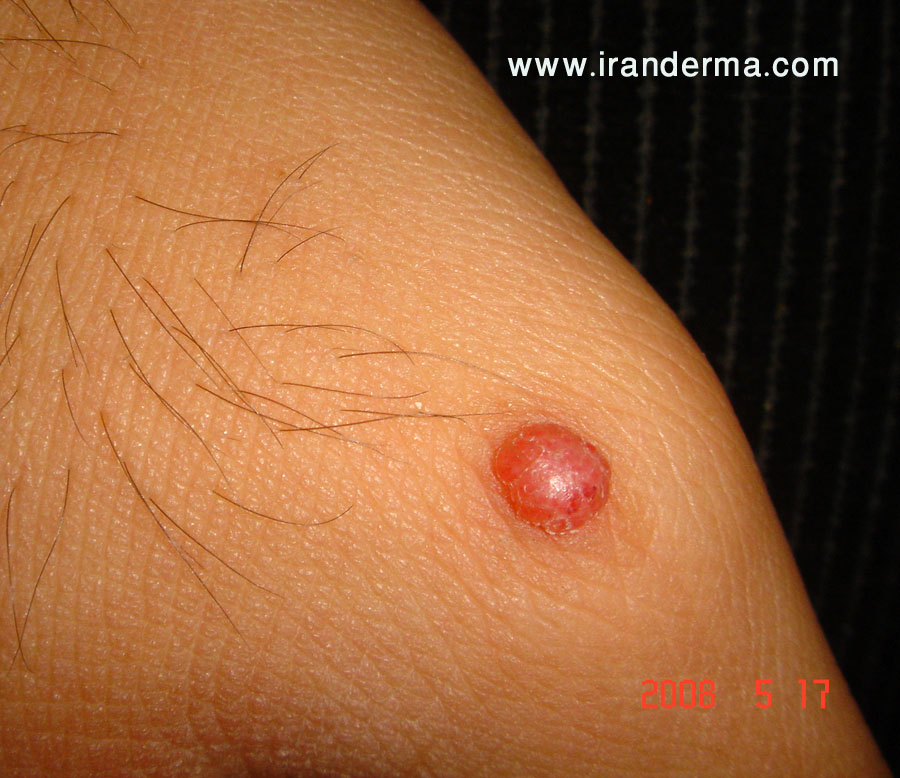
What is your diagnosis for this 20-year-old man?
He had this solitary asymptomatic papule on his hand since about 2 months earlier. No any other significant findings....

The histopathological examination revealed a tumor composed of mixed epithelioid and spindle cells as nests and small clusters. Now, making the diagnosis is much easier!
Diagnosis: Spitz Nevus
| From
eMedicine.com: The classic Spitz nevus is predominantly compound, although junctional and intradermal lesions are also observed. The sine qua non of the diagnosis is the presence of large and/or spindle-shaped melanocytes, usually in nests. The nests are composed of an admixture of spindle cells and/or epithelioid cells, although frequently, the spindle-shaped cells predominate. The spindle cells are usually observed in a fascicular arrangement. These cells have abundant cytoplasm and contain a vesicular nucleus with a conspicuous nucleolus. The epithelioid cells are bizarrely shaped, show poor cohesion, often have several nuclei, and frequently have multiple large nucleoli. Striking symmetry, sharp lateral demarcation, absent (or rare) mitoses, absence of atypical mitoses, presence of eosinophilic and periodic acid-Schiff (PAS)–positive globules (Kamino bodies) and nondisruptive (single-file like) infiltration of collagen are important features indicating the diagnosis of Spitz nevi. Single-file melanocytes may also be observed in the reticular dermis located at the base of the lesion (dispersion). Another important feature is the maturation of cellular elements toward the dermis. Pagetoid spread of the melanocytes is usually confined to the center of the lesion; when present, it can cause confusion with melanoma. The epidermis is hyperkeratotic and acanthotic. A cleavage artifact of fixation is commonly noticed above the nests and around superficial dermal elements. The histologic distinction between Spitz nevi and melanomas is equivocal in up to 8% of cases. |
ايران درما |