IRANDERMA
|
| Home | Medical news | Membership | Cases |
| Selected papers | Text | Dermcalender | Letters/Literature |
| Webwatch | Quiz | Members | SkinCare |
|
Cases |
| Cases 1 to 10 | Cases 11 to 20 | Cases 21 to 30 | Cases 31 to 40 |
Case 4: A well defined nodule on the Lt.buttock of a 2 yrs old baby...
Case 5: A 31 yrs old man with disseminated annular lesions and...
Case 6: Multiple purple nodules and plaques on the face and back ...
Case 7: Painful non-healing ulcer following a trivial trauma...
Case 8: A 11 years old girl with 4 months history for an asymptomatic linear ...
Case 9: Pruritic pigmented plaques on the trunk and proximal extremities of ..
Case 10: A 42 yrs old healthy woman presented with a 2 yrs history of multiple..
Case 1:Can you guess what was the
skin disease of WC?
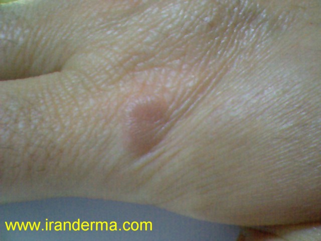
Case 2:A 48 years old female presented with palmar nodule on the ulnar half of her Lt.hand and flexion contracture of her Rt.hand. What is your diagnosis and do you know what are the associated disorders to this disease?

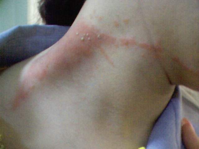
Case 3:A 29 years old female with acute onset of a linear erythematous vesiculopustular eruption on her neck. What is your diagnosis?!
Case 4: A 2 years old boy referred for a yellowish nodular lesion on his Lt.buttock since about 3 months ago. In PE, the nodule was firm and rubbery and a small satellite papules seen in the periphery of the nodule. Otherwise the patient was normal. The lesion totally excised; histopathologic examination showed a diffuse infiltration of histiocytoid cell, admixed with spindle cells and scattered Touton and foreign body giant cells. What's your diagnosis ?

Case 5: A 31 years old man referred with disseminated annular lesions on the trunk and extremities. In PE thinned and lost eyelashes were remarkable. What's your diagnosis?
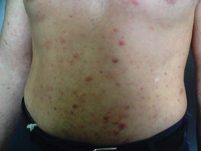
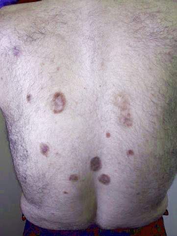
Case 6:
A 40-year-old man presented with multiple purple plaques and nodules,
which had been present on the back for approximately 3 years.
The lesions had gradually extended over the face, trunk and proximal
extremities. He had no symptom except occasional mild pruritus. The patient was
in good health and was on no medications. Physical
examinations revealed multiple violaceous to brown, indurated, 5-50 mm, round to
oval plaques on the face, arms, shoulders, and back, as well as a solitary
lesion on the right thigh. Surface telangiectases were noted, especially on the
shoulder lesions. There was no scaling or ulceration. Routine laboratory tests
were unremarkable.
Histopathology revealed a diffuse, relatively dense infiltrate of neutrophils, eosinophils and mononuclear inflammatory cells in dermis with an obvious Grenz zone.
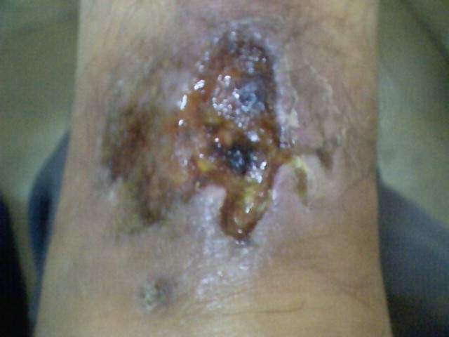
Case 7: A 26 years old man referred for a painful ulcer on his left lower leg which started initially following a trivial trauma since 5 months ago. A skin biopsy from the border of the lesion revealed massive neutrophilic infiltration, plus necrosis of the vascular wall, extravasation of erythrocytes and swelling of the endothelial cells.
Case 8: A 11 years old girl with 4 months history for an asymptomatic linear papular lesion on her left forearm. Skin biopsy revealed a moderately dense lymphohistiocytic infiltrate on the upper dermis and also around the subpapillary vessels with parakeratosis and slight acanthosis.

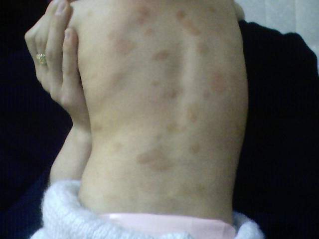
Case 9: A 2 yrs old girl presented with pruritic pigmented plaques over her trunk and proximal extremities since 6 months of age. The histopathological examination was diagnostic .
Case 10: A 42 yrs old healthy woman presented with a 2 yrs history of multiple papules on her hands. The lesions had been mildly itchy. Physical examination revealed discrete erythematous papules distributed over back of her hands and lateral aspects of fingers. The histopathological examination was diagnostic.