|
Quiz: December 2004 |
A 65-year-old man presented with a rapidly enlarging papule on the nose since 3 months ago..
What is your diagnosis?
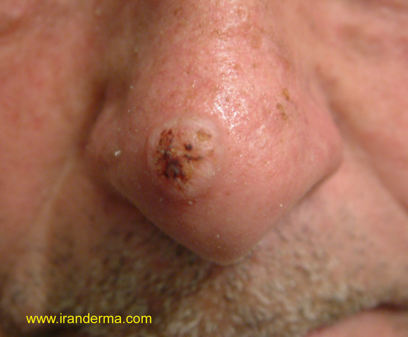
Diagnosis: Keratoacanthoma (KA)
Comment by: M. Mehravaran, MD /Szeged-Hungary
Synonyms:
Self-healing primary squamous carcinoma, molluscum sebaceum, benign
keratoacanthoma and idiopathic cutaneous pseudoepitheliomatus heperplasia.
Definition:
This term describes a suddenly (rapidly) appearing epidermal tumor with
some of the characteristics of an Suamous Cell Carcinoma (SCC) but which
resolves after a short period. Classically considered a benign epithelial
neoplasm, KA shares many clinical and histologic features with SCC, and
indeed, some consider it to be a form of SCC that usually, but not
invarialbly, involutes.
General Features: KAs typically arise on sun-damaged, hair bearing,
light-colored skin in mid-to late life. Males are much more common
affected that females. Lesions have been rarely noted to occur on mucous
membranes and other non-hair-bearing skin such as the lips, intraorally,
and subungually. While usually painless, KAs in certain locations, such
subungual and intraoral, are painful.
The
most interesting feature of this disease is the rapid growth for some 2-6
weeks, which is followed by a stationary period for anther 2-6 weeks, and
finally a spontaneous involution for another 2-6 weeks to leave a slightly
depressed scar. The stationary period and involuting phase are variable ,
some lesions may take six months to a year to completely resolve. It has
been estimated that some 5%
of treated lesions recur, and 10-15% progress to SCC.
CLASSIFICATION
Since
1889, Hutchinson1 report for the first time what now is known as
keratoacanthoma and he designated it "crateriform ulcer of the face.
There are many types of KAs.
·
Solitary
(Hutchinson) KA.
This type is a rapidly growing papule, which enlarges from a 1-mm
macule or papule to as much as 25 mm in 3-8 weeks. When fully
developed it is a hemispheric, dome-shaped, skin-colored nodule in
which there is a smooth crater filled with a central keratin plug. The
smooth shiny lesion is sharply demarcated from it surroundings.
Telangiectases may run through the lesions. The solitary KA occurs
mostly on sun-exposed skin, with the central portion of the face, the
backs of the hands, and the arms being the most commonly involved sites.
Rarley this tumor involves the mucous membranes of the oral cavity or
subungual region. KA is seen mostly in middle-aged to elderly persons,
with men being more frequently involved. Subungual lesions are
painful and induce early underlying bony destruction, characterized on
X-ray as a crescent-shaped lytic defect without accompanying sclerosis of
perosteal reaction.
·
Giant KA. Is
a rapidly progressive lesion with growth up to 3 cm in diameter. The bulk
of such neoplasms accounts for their significant local destractiveness.
·
KA centrifugum
marginatum. Is
characterized by rapidly enlarging plaque with central clearing, resulting
in an annular configuration.
·
Mutiple
“self_healing” KA (Ferguson-Smith) type of KA are
characterized by generalized lesions, which often ulcerate. The condition
is inherited (autosomal dominant) trait.
· Another rare presentation is generalized eruptive (Grzybowski) KA. The lesions in this type typically occur in adulthood and may number in the thousands. Systemic retinoids, both isotretinoin and etretinate, have been used successfully in treating multiple keratoacanthoma. Eruptive KA is characterized by a generalized eruption of numerous dome-shaped, skin-colored papules from 2-7 mm in diameter. The eruption is usually generalized but spares the palms and soles.
KAs in certain
locations, such subungual and intraoral, are painful.
·
KAs have also been
described as occurring in increased number in patients with xeroderma
pigmentosum, Torre’s Syndrome, and in renal transplant (or other
immunsuppressed) patients.
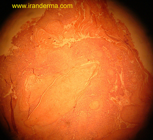
Pathology:
KA has a characteristic symmetrical cup of flask-shaped structure. There
is a minor degree of
epidermal dysplasia and little evidence of tissue invasion by the
epithelium (cup-shaped epidermal invagination)
KA
is most readily diagnosed at scanning magnification of an optimally
sectioned specimen because of its distinctive architecture. It is an
exo-endophytic proliferation of cornifying squamous epithelium having a
central crater filled with orthokeratotic scale. The overall lesion is
symmetrical, but confusion often arises in tangentially sectioned
specimens or incomplete biopsy specimens because the characteristic flask-
or cup-shaped architecture cannot be recognized.
According
to Dr. Bernard Ackerman.
Keratoacanthomas are a special type of squamous-cell carcinoma. The type called "solitary" (but which may be multiple) occurs on skin injured by the effects of sunlight and is characterized by rapid growth and often by involution in months in the absence of therapy. It has a characteristic crateriform appearance as a consequence of dilation of contiguous infundibula that are filled with corneocytes. The squamous-cell carcinoma seems to derive from infundibular keratinocytes. The "solitary" kind of KA is a squamous-cell carcinoma, not only for reasons that pertain to architectural and cytologic characteristics, but because, rarely, the neoplasm metastasizes (especially in persons who are immunosuppressed).
Subungual KA is wholly unrelated to "solitary" KA, being unassociated as it is with follicles. It also has a crateriform appearance and it burrows, slowly but surely, through the subungual soft tissues and often into bone of the distal phalanx. Other types of KA are the eruptive, the familial, and the gigantic.
Treatment: Excision or curettage and fulguration. The intralesional
injection of triamcinolone or 5-FU.
Belomycin. Oral etretinate, Isotretinoin. X-Ray Therapy.
References :
1. Hutchinson J. The crateriform ulcer of the face: A form of acute epithelial cancer. Trans Pathol Soc London 1889;40: 275.
![]()
ايران درما |