IRANDERMA |
|
Quiz: July 2007 |
What is your diagnosis for this Young lady?
She has had this solitary lesion on the volar aspect of her thumb since months ago. It was mildly tender. She had no significant findings in past medical history and general examinations.
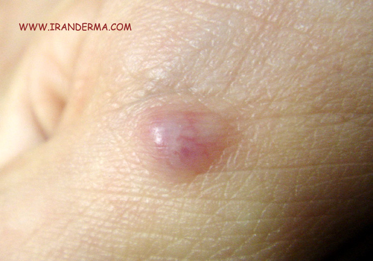
Diagnosis: Glomus Tumor
Omid Zargari;
The glomus tumor, first described by Masson's in 1924, is a relatively rare hamartoma derived from a glomus body and clinically presents usually as a solitary tumor. As many of you correctly mentioned angiomas, angioleiomyoma, Spitz nevus, malignant melanoma and myxoid cyst could be considered as differential for this case. Below, please read comments by our readers;
1.Eccrine poroma; but this lesion has often a scaly/keratotic collar. It arises from eccrine duct epithelium & has a malignancy potential.
2.Myxoid cyst (mucinous cust): it is seen mostly over dorsal aspects of DIP joints.
3.Glomus tumor:painful tumor of Sucquet Hoyer canal.
Glomus tumors arise from the arterial portion of the glomus body, or the Sucquet-Hoyer canal, which is an arteriovenous shunt in the dermis that contributes to temperature regulation. Although glomus tumors are thought to arise from glomus cells, these tumors have been observed in extracutaneous locations that are not known to contain glomus cells. One explanation for this finding is that these tumors may arise from perivascular cells that can differentiate into glomus cells. Multiple glomus tumors, especially the disseminated variant, are inherited in an autosomal-dominant pattern with incomplete penetrance.
The exact incidence of glomus tumors is unknown. The multiple variant is rare, accounting for less than 10% of all cases. The probable misdiagnosis of many of these lesions as hemangiomas or venous malformations also makes an accurate assessment of incidence difficult.
- Patients with solitary glomus tumors usually have paroxysmal pain, which can be severe and exacerbated by pressure or temperature changes, especially cold.
- Multiple glomus tumors can also be painful, but this feature is less common, and the pain usually is not severe.
- Patients with multiple lesions often seek medical attention because they are worried or have cosmetic concerns.
- Because multiple glomus
tumors are inherited as an
autosomal-dominant condition, a
family history of similar
lesions may be helpful for
diagnosis.
- Solitary glomus tumors have the following characteristics:
-
- Blue or purple
- Papules or nodules that can be blanched
- Size usually smaller than 1 cm
- Located most commonly in acral areas, especially subungual areas of fingers and toes
- The multiple variant is subdivided into regional or localized, disseminated, and congenital plaquelike forms.
-
- The regional variant consists of blue-to-purple partially compressible papules or nodules that are grouped and limited to a specific area, most commonly an extremity.
- The disseminated type consists of multiple lesions distributed over the body with no specific grouping. This form is less common than the regional variant.
- Congenital plaquelike glomus tumors consist of either grouped papules that coalesce into indurated plaques or clusters of discrete nodules. This form is the rarest variant of multiple glomus tumors.
- Two useful items for diagnosing glomus tumors, particularly solitary painful glomus tumors (especially those under a nail) are the following:
-
- Hildreth sign, which is disappearance of pain after application of a tourniquet proximally on the arm
- Love test, which consists of eliciting pain by applying pressure to a precise area with the tip of a pencil
- Features of
glomangiosarcomas may
include the following:
- Size larger than 1 cm
- Rapid growth
- Deep soft tissue
involvement
- Glomus tumors are neoplasms caused by a proliferation of glomus cells, which make up a portion of the glomus body.
- The initiating event for glomus cell proliferation is unknown.
- Some authors have postulated that trauma induces solitary subungual glomus tumors, although this theory is not well studied.
- Most multiple glomus tumors, especially those of the disseminated form, are inherited in an autosomal dominant pattern with incomplete penetrance. Most hereditary glomangiomas are associated with defects in the glomulin gene, located on chromosome 1.
-
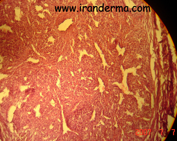
Histologic Findings
Solitary and multiple glomus tumors have distinct histopathologic features.Solitary lesions appear mostly as solid well-circumscribed nodules surrounded by a rim of fibrous tissue. They contain endothelium-lined vascular spaces surrounded by clusters of glomus cells. The glomus cells are monomorphous round or polygonal cells with plump nuclei and scant eosinophilic cytoplasm.Multiple lesions are less well circumscribed and less solid-appearing than their solitary counterparts. Multiple lesions have the overall appearance of a hemangioma. They contain multiple irregular, dilated, endothelium-lined vascular channels that contain red blood cells. The vascular spaces are larger than those in solitary glomus tumors. Small aggregates of glomus cells are present in the walls of these channels and in small clusters in the adjacent stroma. Multiple glomus tumors have more narrow and focal aggregates of glomus cells than solitary lesions. The overall appearance of multiple glomus tumors accounts for their alternate name, glomangiomas.Glomangiosarcomas resemble benign glomus tumors. However, glomangiosarcomas have more atypia, pleomorphism, and mitotic figures, and they have an invasive growth pattern. Most often, foci of benign glomus tumor are correlated with malignant lesions. -
Treatment:
-
Surgical Care
- The treatment of choice for solitary glomus tumors is surgical excision.
-
- For multiple glomus tumors, excision may be more difficult because of their poor circumscription and the large number of lesions.
- Excision should be limited to symptomatic lesions.
- Other reported treatment modalities include argon and carbon dioxide laser therapy and sclerotherapy with hypertonic saline or sodium tetradecyl sulfate. These are most useful in treating multiple lesions.
- The treatment of glomangiosarcoma is based on a few case reports.
-
- Wide local excision is adequate treatment and probably the treatment of choice.
- However, geometric excision is probably a reasonable alternative in cosmetically sensitive areas
Prognosis
- The prognosis is excellent.
- Excision of painful lesions most often results in cure, with a low recurrence rate for solitary lesions.
- Malignant glomus tumors are extremely rare and usually locally aggressive.
- Their overall prognosis is good when they are treated with wide excision. However, metastases do occur and are associated with a very poor prognosis.
![]()
ايران درما |