IRANDERMA |
|
Quiz: September 2007 |
What is your diagnosis?

Diagnosis: Verrucous Hemangioma
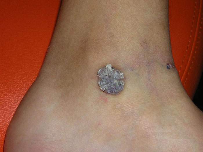
Verrucous hemangioma (VH) commonly presents at birth although it may appear later in life. Lesions can be single or grouped, sometimes associated with small satellite lesions, and they occur mostly on the legs.
VH starts as well-defined, macular areas of vascular staining resembling port-wine stains that with time it takes the characteristic bluish hue and a verrucous surface.
Histologically, VH is characterized by a hyperplastic epidermis and the underlying dermis consisting of lobules of capillaries and vascular spaces.
Clinically VH can be misdiagnosed as infantile hemangioma, angiokeratoma, and rarely as lymphangioma circumscriptum.
VH is best treated by excision, but there is a tendency to recur unless excision is complete.
Shahriar Nazari, Istanbul/ Turkey
ايران درما |